



Upper Root Tip Forceps: Selection Guide for Dentists
- , by SurgiMac
- 26 min reading time
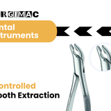
Learn how upper root tip forceps support maxillary root removal, with practical guidance on beak design, access, anatomy, selection, and reprocessing.
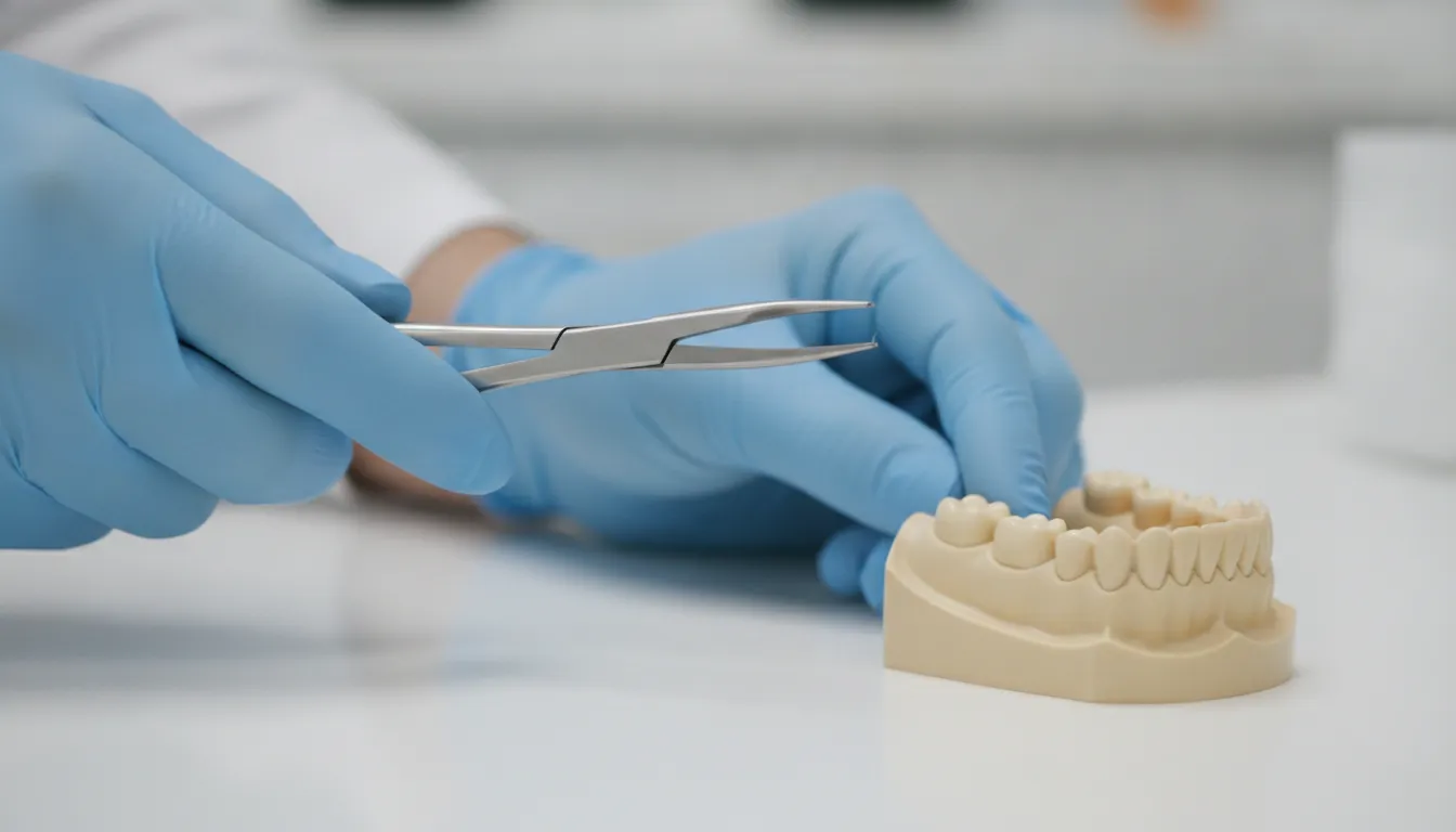
Upper root tip forceps help dental professionals approach small maxillary root remnants with a focused beak profile and controlled hand position.
Review precision dental instruments from SurgiMac
Upper root tip forceps are specialized extraction instruments used to grasp small root remnants or fractured tips that conventional crown forceps cannot securely engage. For maxillary cases, selection should account for fragment depth, root morphology, visibility, access, beak shape, and the direction of controlled movement.
Removing a fractured or retained root tip in the maxillary arch demands more than a narrow instrument. Posterior access may be restricted by the cheek, adjacent teeth, the curvature of the upper arch, or limited visibility. Root anatomy also varies by tooth and patient. The forceps should support the operator's planned approach, not replace radiographic assessment, tissue management, illumination, suction, or appropriate surgical judgment.
This guide explains the clinical role of upper root tip forceps, the design features that affect access and control, and a practical way to select and maintain them for professional dental use.
What Are Upper Root Tip Forceps Used For?
Upper root tip forceps are intended for small, accessible root remnants or fractured tips that lack enough coronal structure for routine crown forceps. Their narrow working ends can provide a more deliberate purchase, while the clinician still evaluates whether the fragment is visible, mobile, and safely accessible.
Removing fractured and retained root fragments
A root may separate during extraction, leaving a fragment that cannot be engaged effectively with a broad crown forceps. A residual root can also remain after the crown has been removed or become structurally unavailable for conventional purchase. In these situations, a specialized root instrument may help the operator approach the fragment with less unnecessary contact around the socket.
The instrument is not selected solely because the fragment is small. The clinician should consider its location, orientation, mobility, surrounding bone, and relationship to nearby structures. A visible coronal portion that can be approached along a controlled path is a different problem from a deep, immobile, or poorly visualized fragment. The latter may require a different surgical plan rather than progressively greater force.
The NCBI Bookshelf reference on dental extraction instruments provides useful background on forceps and root-fragment removal: review the dental extraction instrument reference.
Why the upper arch changes the approach
Maxillary root removal can involve a curved working field and restricted posterior access. An offset or bayonet-style shank may position the handle away from the operator's direct sightline and create useful hand clearance. The design can make it easier to align the beaks with the fragment while maintaining a stable wrist and a clear view of the working end.
These features do not remove the need to assess the maxillary sinus, adjacent teeth, alveolar bone, or the risk of fragment displacement. They simply give the operator another way to manage the geometry of the approach. The right instrument is the one that fits the anatomy and planned movement, not necessarily the one with the narrowest beaks.
When general extraction forceps may be unsuitable
Broad crown forceps are useful when enough tooth structure remains for the beaks to engage securely. On a narrow apical remnant, however, a broad instrument may offer an unstable purchase or contact tissue and adjacent tooth structure unnecessarily. Upper root tip forceps are better considered as one component of a broader extraction set that may include elevators, luxators, surgical burs, curettes, and other instruments selected for the case.
For related guidance on maxillary extraction access and instrument selection, see SurgiMac's maxillary molar forceps guide. Practices comparing focused extraction tools can also review the Titanium Black Series for instrument options designed to support visibility during delicate work.
How Does Maxillary Design Improve Root Tip Access?
Maxillary root fragments can be difficult to approach because the posterior upper arch curves away from a direct line of sight. An upper root tip forceps with narrow, curved beaks and an offset or bayonet shank can help the clinician reach an accessible fragment without forcing the hand into an awkward position.
Beak shape and working angle
Beak geometry determines how the instrument approaches the fragment and where contact occurs. Narrow beaks can enter a confined space, but they must still be seated deliberately. Curved beaks may follow the contour of the upper arch and help maintain a more useful line of sight. An angled working end may also keep the handle clear of adjacent teeth or soft tissue.
These design features should be evaluated together. A narrow beak with an unsuitable angle may be difficult to control. A sharply curved profile may improve access in one posterior position but be less suitable in another. The operator should inspect the beaks, compare the working end with the expected fragment location, and confirm that the design supports the intended direction of movement.
Offset and bayonet profiles
Offset and bayonet configurations can help separate the handle from the line between the operator's hand and the root fragment. That separation may improve visibility and provide more room around the cheek, neighboring teeth, or a limited posterior opening. It can also support a more neutral wrist position during a focused maneuver.
Better access does not mean the forceps should be used with greater force. A clear sightline should make it easier to confirm beak placement, avoid soft tissue engagement, and stop when the purchase is not secure. If the fragment cannot be visualized or the instrument cannot be seated safely, the clinician should reassess the approach rather than repeatedly loading the forceps.
| Design feature | Potential access benefit | Control consideration |
|---|---|---|
| Narrow beaks | Reach a small, accessible root remnant within a restricted space. | Seat the tips carefully and avoid soft tissue or adjacent tooth contact. |
| Curved beaks | Follow the contour of the upper arch and support posterior access. | Confirm that the curve matches the fragment position and planned movement. |
| Offset or bayonet shank | Move the handle away from the sightline and improve hand clearance. | Maintain a stable grip and verify the working end remains visible. |
| Serrated or textured beak surfaces | Support purchase when the instrument and fragment are appropriately matched. | Use controlled pressure and follow the manufacturer's intended use. |
How Should Clinicians Select Upper Root Tip Forceps?
Select upper root tip forceps by matching the instrument to the fragment's anatomy, depth, visibility, access path, and expected direction of controlled movement. A structured selection process helps the dental team avoid choosing an instrument by name alone or applying a narrow forceps to a case that requires a different approach.
Assess fragment location and visibility
Start by determining where the fragment is located and whether it can be seen directly or approached predictably. The team should review the available clinical and radiographic information, identify relevant adjacent structures, and consider whether the fragment is coronal enough for the selected beaks to engage. Visibility is especially important in the posterior maxilla, where the working field can be difficult to inspect.
A forceps that appears appropriate on a tray may not be appropriate once the patient position, cheek retraction, opening, and tooth position are considered. Select for the actual working field rather than a catalog label.
Match beaks to root morphology
Root shape and remaining purchase influence the working end that is most likely to provide stable contact. Consider whether the remnant is slender, broad, curved, fractured irregularly, or positioned at an angle. The beaks should be capable of reaching the accessible portion without requiring the operator to twist the instrument or use an unstable hand position.
Do not confuse a smaller beak with a safer beak. The goal is a controlled, visible purchase that fits the fragment. If the beaks contact only a sharp edge or cannot be seated along a stable surface, the instrument may not be suitable.
Evaluate hand position and access
Compare straight, curved, offset, or bayonet profiles with the expected path of access. The handle should allow the operator to maintain a steady grip and useful wrist position while keeping the beaks in view. In a posterior maxillary site, an offset profile may improve clearance. In another location, a different orientation may offer better control.
Confirm the product specification
Before adding an instrument to a clinical set, confirm its product description, intended application, materials, dimensions, and manufacturer instructions. SurgiMac's upper-root extracting forceps product page is one example of a product-specific reference for upper-root instrumentation. Product details should be evaluated alongside the patient's anatomy and the clinician's technique.
- Identify the fragment. Review its location, visibility, morphology, mobility, and surrounding anatomy.
- Define the access path. Determine the direction that offers the clearest view and safest hand position.
- Compare working ends. Match beak width, curvature, angle, and surface profile to the available purchase.
- Check clearance. Confirm that the handle and shank can pass without unnecessary contact with adjacent structures.
- Test the planned seating mentally. Ensure the beaks can engage the fragment without relying on a twisting or unstable movement.
- Stop and reassess when the purchase is poor. A specialized forceps does not make an inaccessible or unsafe fragment suitable for closed engagement.
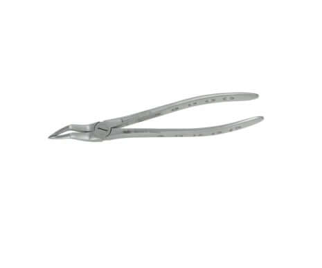
Review upper-root extraction forceps for your instrument set
Upper Root Tip Forceps vs. General Extraction Forceps
Upper root tip forceps and general extraction forceps serve different access problems. General forceps often provide broader beaks for engaging remaining crown structure, while root tip instruments emphasize access to a small remnant. The correct choice depends on the available purchase and anatomy, not on the assumption that one instrument is universally preferable.
| Selection question | Upper root tip forceps | General extraction forceps |
|---|---|---|
| What structure is available? | A small, accessible root remnant or fractured tip. | A crown or larger portion of tooth structure suitable for broader beaks. |
| What access is needed? | Focused access around a narrow fragment, often in a restricted field. | Broader access around a tooth with more available purchase. |
| What should guide selection? | Fragment depth, beak fit, visibility, root morphology, and hand clearance. | Tooth anatomy, crown form, arch position, and intended extraction movement. |
Using a broad forceps on a small remnant can create an unstable purchase. Using a root tip forceps when the fragment is deep, poorly visualized, or inaccessible can also be inappropriate. The instrument should be selected after the operator defines the access problem and the limits of controlled engagement.
Which Instrument Features Support Precision?
Precision depends on the interaction of instrument geometry, grip, visibility, and operator technique. Brand series can help a practice organize its instrument options, but a series name should never replace evaluation of the individual working end and the clinical task.
Air Series for balance and control
SurgiMac's Air Series is positioned around comfort, control, and precision. Those priorities may be relevant when a clinician needs steady handling during a focused root-removal workflow. Review the individual product specifications and compare the handle, balance, and working end with the planned maxillary access path.
Slim Series for compact access
The Slim Series emphasizes compact ergonomic instrument design. A compact profile may be useful when a clinician is comparing options for hand clearance or a confined working field. It should still be assessed against the root's location, the available sightline, and the beak geometry required for secure engagement.
Titanium Black Series for reduced glare
The Titanium Black Series combines a dark surface treatment with surgical and dental instrument functionality. Reduced surface glare may be helpful in delicate work, depending on the clinical environment and lighting. As with any instrument, the finish does not determine whether the beaks fit the root fragment. Selection remains anatomy- and task-specific.
For a dental practice, these lines can support different preferences for balance, handle profile, visibility, and workflow organization. The final choice should be based on the actual instrument specification, reprocessing requirements, and clinical use case.
How Should Dental Teams Inspect and Reprocess Them?
Dental teams should inspect, clean, sterilize, and store upper root tip forceps according to the manufacturer's instructions, the practice infection-control plan, and applicable regulatory guidance. Reprocessing is part of instrument performance because residue, corrosion, damaged beaks, or a compromised hinge can affect both function and patient safety.
Inspect before and after use
Before use, check the beaks for alignment, chips, deformation, corrosion, residue, or other damage. Open and close the instrument as appropriate to confirm smooth movement. After use, remove gross soil promptly according to the practice protocol and prevent contaminated instruments from drying when the validated workflow calls for an immediate pre-cleaning step.
Follow validated cleaning and sterilization steps
Use the cleaning method, detergents, instrument trays, lubrication steps, packaging materials, and sterilization cycle specified by the manufacturer and the practice's validated process. Do not assume that a general metal instrument procedure is suitable for every design or finish. The CDC's summary of sterilization and disinfection practices for dental settings provides a useful reference: review CDC dental infection-control guidance.
Remove damaged instruments from service
If the beaks no longer align, the hinge binds, the surface is damaged, or the instrument cannot be cleaned effectively, remove it from service for evaluation. A forceps that does not close or seat predictably may reduce control during a procedure. Store clean, dry, processed instruments in a way that protects the working ends and supports traceability within the practice workflow.
Explore SurgiMac Slim Series instruments for precision-focused workflows
Frequently Asked Questions
What are upper root tip forceps used for?
Upper root tip forceps are used to grasp small, accessible root remnants or fractured tips that do not provide enough structure for broad crown forceps. The clinician should confirm visibility, beak fit, fragment position, and surrounding anatomy before choosing them.
How does maxillary design improve access to upper root fragments?
Curved beaks and offset or bayonet shanks can follow the upper arch and move the handle away from the direct sightline. This may improve hand clearance and visibility in posterior areas, but it does not replace radiographic assessment or careful surgical judgment.
How should clinicians select the right instrument for a maxillary root tip?
Match the beak width, curvature, angle, and surface profile to the fragment's morphology, depth, visibility, and available access. Also assess wrist position, handle clearance, the planned direction of movement, and the manufacturer's product specifications.
How should upper root tip forceps be maintained between procedures?
Inspect the beaks and hinge, remove soil promptly, and follow the manufacturer's validated cleaning, packaging, sterilization, and storage instructions. Remove instruments with damaged surfaces, misaligned beaks, binding hinges, or any defect that affects predictable function.
Upper root tip forceps help dental professionals approach small maxillary root remnants with a focused beak profile and controlled hand position.
Review precision dental instruments from SurgiMac
Upper root tip forceps are specialized extraction instruments used to grasp small root remnants or fractured tips that conventional crown forceps cannot securely engage. For maxillary cases, selection should account for fragment depth, root morphology, visibility, access, beak shape, and the direction of controlled movement.
Removing a fractured or retained root tip in the maxillary arch demands more than a narrow instrument. Posterior access may be restricted by the cheek, adjacent teeth, the curvature of the upper arch, or limited visibility. Root anatomy also varies by tooth and patient. The forceps should support the operator's planned approach, not replace radiographic assessment, tissue management, illumination, suction, or appropriate surgical judgment.
This guide explains the clinical role of upper root tip forceps, the design features that affect access and control, and a practical way to select and maintain them for professional dental use.
What Are Upper Root Tip Forceps Used For?
Upper root tip forceps are intended for small, accessible root remnants or fractured tips that lack enough coronal structure for routine crown forceps. Their narrow working ends can provide a more deliberate purchase, while the clinician still evaluates whether the fragment is visible, mobile, and safely accessible.
Removing fractured and retained root fragments
A root may separate during extraction, leaving a fragment that cannot be engaged effectively with a broad crown forceps. A residual root can also remain after the crown has been removed or become structurally unavailable for conventional purchase. In these situations, a specialized root instrument may help the operator approach the fragment with less unnecessary contact around the socket.
The instrument is not selected solely because the fragment is small. The clinician should consider its location, orientation, mobility, surrounding bone, and relationship to nearby structures. A visible coronal portion that can be approached along a controlled path is a different problem from a deep, immobile, or poorly visualized fragment. The latter may require a different surgical plan rather than progressively greater force.
The NCBI Bookshelf reference on dental extraction instruments provides useful background on forceps and root-fragment removal: review the dental extraction instrument reference.
Why the upper arch changes the approach
Maxillary root removal can involve a curved working field and restricted posterior access. An offset or bayonet-style shank may position the handle away from the operator's direct sightline and create useful hand clearance. The design can make it easier to align the beaks with the fragment while maintaining a stable wrist and a clear view of the working end.
These features do not remove the need to assess the maxillary sinus, adjacent teeth, alveolar bone, or the risk of fragment displacement. They simply give the operator another way to manage the geometry of the approach. The right instrument is the one that fits the anatomy and planned movement, not necessarily the one with the narrowest beaks.
When general extraction forceps may be unsuitable
Broad crown forceps are useful when enough tooth structure remains for the beaks to engage securely. On a narrow apical remnant, however, a broad instrument may offer an unstable purchase or contact tissue and adjacent tooth structure unnecessarily. Upper root tip forceps are better considered as one component of a broader extraction set that may include elevators, luxators, surgical burs, curettes, and other instruments selected for the case.
For related guidance on maxillary extraction access and instrument selection, see SurgiMac's maxillary molar forceps guide. Practices comparing focused extraction tools can also review the Titanium Black Series for instrument options designed to support visibility during delicate work.
How Does Maxillary Design Improve Root Tip Access?
Maxillary root fragments can be difficult to approach because the posterior upper arch curves away from a direct line of sight. An upper root tip forceps with narrow, curved beaks and an offset or bayonet shank can help the clinician reach an accessible fragment without forcing the hand into an awkward position.
Beak shape and working angle
Beak geometry determines how the instrument approaches the fragment and where contact occurs. Narrow beaks can enter a confined space, but they must still be seated deliberately. Curved beaks may follow the contour of the upper arch and help maintain a more useful line of sight. An angled working end may also keep the handle clear of adjacent teeth or soft tissue.
These design features should be evaluated together. A narrow beak with an unsuitable angle may be difficult to control. A sharply curved profile may improve access in one posterior position but be less suitable in another. The operator should inspect the beaks, compare the working end with the expected fragment location, and confirm that the design supports the intended direction of movement.
Offset and bayonet profiles
Offset and bayonet configurations can help separate the handle from the line between the operator's hand and the root fragment. That separation may improve visibility and provide more room around the cheek, neighboring teeth, or a limited posterior opening. It can also support a more neutral wrist position during a focused maneuver.
Better access does not mean the forceps should be used with greater force. A clear sightline should make it easier to confirm beak placement, avoid soft tissue engagement, and stop when the purchase is not secure. If the fragment cannot be visualized or the instrument cannot be seated safely, the clinician should reassess the approach rather than repeatedly loading the forceps.
| Design feature | Potential access benefit | Control consideration |
|---|---|---|
| Narrow beaks | Reach a small, accessible root remnant within a restricted space. | Seat the tips carefully and avoid soft tissue or adjacent tooth contact. |
| Curved beaks | Follow the contour of the upper arch and support posterior access. | Confirm that the curve matches the fragment position and planned movement. |
| Offset or bayonet shank | Move the handle away from the sightline and improve hand clearance. | Maintain a stable grip and verify the working end remains visible. |
| Serrated or textured beak surfaces | Support purchase when the instrument and fragment are appropriately matched. | Use controlled pressure and follow the manufacturer's intended use. |
How Should Clinicians Select Upper Root Tip Forceps?
Select upper root tip forceps by matching the instrument to the fragment's anatomy, depth, visibility, access path, and expected direction of controlled movement. A structured selection process helps the dental team avoid choosing an instrument by name alone or applying a narrow forceps to a case that requires a different approach.
Assess fragment location and visibility
Start by determining where the fragment is located and whether it can be seen directly or approached predictably. The team should review the available clinical and radiographic information, identify relevant adjacent structures, and consider whether the fragment is coronal enough for the selected beaks to engage. Visibility is especially important in the posterior maxilla, where the working field can be difficult to inspect.
A forceps that appears appropriate on a tray may not be appropriate once the patient position, cheek retraction, opening, and tooth position are considered. Select for the actual working field rather than a catalog label.
Match beaks to root morphology
Root shape and remaining purchase influence the working end that is most likely to provide stable contact. Consider whether the remnant is slender, broad, curved, fractured irregularly, or positioned at an angle. The beaks should be capable of reaching the accessible portion without requiring the operator to twist the instrument or use an unstable hand position.
Do not confuse a smaller beak with a safer beak. The goal is a controlled, visible purchase that fits the fragment. If the beaks contact only a sharp edge or cannot be seated along a stable surface, the instrument may not be suitable.
Evaluate hand position and access
Compare straight, curved, offset, or bayonet profiles with the expected path of access. The handle should allow the operator to maintain a steady grip and useful wrist position while keeping the beaks in view. In a posterior maxillary site, an offset profile may improve clearance. In another location, a different orientation may offer better control.
Confirm the product specification
Before adding an instrument to a clinical set, confirm its product description, intended application, materials, dimensions, and manufacturer instructions. SurgiMac's upper-root extracting forceps product page is one example of a product-specific reference for upper-root instrumentation. Product details should be evaluated alongside the patient's anatomy and the clinician's technique.
- Identify the fragment. Review its location, visibility, morphology, mobility, and surrounding anatomy.
- Define the access path. Determine the direction that offers the clearest view and safest hand position.
- Compare working ends. Match beak width, curvature, angle, and surface profile to the available purchase.
- Check clearance. Confirm that the handle and shank can pass without unnecessary contact with adjacent structures.
- Test the planned seating mentally. Ensure the beaks can engage the fragment without relying on a twisting or unstable movement.
- Stop and reassess when the purchase is poor. A specialized forceps does not make an inaccessible or unsafe fragment suitable for closed engagement.
Review upper-root extraction forceps for your instrument set
Upper Root Tip Forceps vs. General Extraction Forceps
Upper root tip forceps and general extraction forceps serve different access problems. General forceps often provide broader beaks for engaging remaining crown structure, while root tip instruments emphasize access to a small remnant. The correct choice depends on the available purchase and anatomy, not on the assumption that one instrument is universally preferable.
| Selection question | Upper root tip forceps | General extraction forceps |
|---|---|---|
| What structure is available? | A small, accessible root remnant or fractured tip. | A crown or larger portion of tooth structure suitable for broader beaks. |
| What access is needed? | Focused access around a narrow fragment, often in a restricted field. | Broader access around a tooth with more available purchase. |
| What should guide selection? | Fragment depth, beak fit, visibility, root morphology, and hand clearance. | Tooth anatomy, crown form, arch position, and intended extraction movement. |
Using a broad forceps on a small remnant can create an unstable purchase. Using a root tip forceps when the fragment is deep, poorly visualized, or inaccessible can also be inappropriate. The instrument should be selected after the operator defines the access problem and the limits of controlled engagement.
Which Instrument Features Support Precision?
Precision depends on the interaction of instrument geometry, grip, visibility, and operator technique. Brand series can help a practice organize its instrument options, but a series name should never replace evaluation of the individual working end and the clinical task.
Air Series for balance and control
SurgiMac's Air Series is positioned around comfort, control, and precision. Those priorities may be relevant when a clinician needs steady handling during a focused root-removal workflow. Review the individual product specifications and compare the handle, balance, and working end with the planned maxillary access path.
Slim Series for compact access
The Slim Series emphasizes compact ergonomic instrument design. A compact profile may be useful when a clinician is comparing options for hand clearance or a confined working field. It should still be assessed against the root's location, the available sightline, and the beak geometry required for secure engagement.
Titanium Black Series for reduced glare
The Titanium Black Series combines a dark surface treatment with surgical and dental instrument functionality. Reduced surface glare may be helpful in delicate work, depending on the clinical environment and lighting. As with any instrument, the finish does not determine whether the beaks fit the root fragment. Selection remains anatomy- and task-specific.
For a dental practice, these lines can support different preferences for balance, handle profile, visibility, and workflow organization. The final choice should be based on the actual instrument specification, reprocessing requirements, and clinical use case.
How Should Dental Teams Inspect and Reprocess Them?
Dental teams should inspect, clean, sterilize, and store upper root tip forceps according to the manufacturer's instructions, the practice infection-control plan, and applicable regulatory guidance. Reprocessing is part of instrument performance because residue, corrosion, damaged beaks, or a compromised hinge can affect both function and patient safety.
Inspect before and after use
Before use, check the beaks for alignment, chips, deformation, corrosion, residue, or other damage. Open and close the instrument as appropriate to confirm smooth movement. After use, remove gross soil promptly according to the practice protocol and prevent contaminated instruments from drying when the validated workflow calls for an immediate pre-cleaning step.
Follow validated cleaning and sterilization steps
Use the cleaning method, detergents, instrument trays, lubrication steps, packaging materials, and sterilization cycle specified by the manufacturer and the practice's validated process. Do not assume that a general metal instrument procedure is suitable for every design or finish. The CDC's summary of sterilization and disinfection practices for dental settings provides a useful reference: review CDC dental infection-control guidance.
Remove damaged instruments from service
If the beaks no longer align, the hinge binds, the surface is damaged, or the instrument cannot be cleaned effectively, remove it from service for evaluation. A forceps that does not close or seat predictably may reduce control during a procedure. Store clean, dry, processed instruments in a way that protects the working ends and supports traceability within the practice workflow.
Explore SurgiMac Slim Series instruments for precision-focused workflows
Frequently Asked Questions
What are upper root tip forceps used for?
Upper root tip forceps are used to grasp small, accessible root remnants or fractured tips that do not provide enough structure for broad crown forceps. The clinician should confirm visibility, beak fit, fragment position, and surrounding anatomy before choosing them.
How does maxillary design improve access to upper root fragments?
Curved beaks and offset or bayonet shanks can follow the upper arch and move the handle away from the direct sightline. This may improve hand clearance and visibility in posterior areas, but it does not replace radiographic assessment or careful surgical judgment.
How should clinicians select the right instrument for a maxillary root tip?
Match the beak width, curvature, angle, and surface profile to the fragment's morphology, depth, visibility, and available access. Also assess wrist position, handle clearance, the planned direction of movement, and the manufacturer's product specifications.
How should upper root tip forceps be maintained between procedures?
Inspect the beaks and hinge, remove soil promptly, and follow the manufacturer's validated cleaning, packaging, sterilization, and storage instructions. Remove instruments with damaged surfaces, misaligned beaks, binding hinges, or any defect that affects predictable function.
Tags
Blog posts
-
, by SurgiMac A Clinician's Guide to Absorbable Suture Material
-
, by SurgiMac A Clinician's Guide to Dental Composite Resin Kits
-

, by SurgiMac How to Disinfect with Metrex CaviWipes Correctly
