A Guide to Bone Graft Material for Dental Implants
- , by SurgiMac
- 45 min reading time
Get a clear overview of bone graft material options for dental implants, including autografts, allografts, xenografts, and synthetic choices.
A dental implant's success starts with a solid foundation in the jawbone. When you recommend bone augmentation, your patient's first question is almost always, "Where does the bone come from?" Your ability to answer this clearly builds immense trust. Explaining the different types of bone graft material—from the patient's own bone to synthetic substitutes—is key. This guide breaks down each bone graft material, giving you the clinical details to choose the right option and communicate it with confidence. This helps your patient feel informed and ready to proceed with treatment.
Key Takeaways
- Prioritize Bone Volume for Implant Success: A successful dental implant requires a solid foundation of adequate bone. Bone grafting is the standard procedure to rebuild this foundation, using one of four material types—autografts, allografts, xenografts, or alloplasts—to ensure long-term stability.
- Match the Graft Material to the Clinical Need: The ideal graft is determined by the specific case. Base your selection on a careful assessment of the defect's size and location, the patient's overall health and healing capacity, and your surgical timeline.
- Understand the Core Clinical Trade-Offs: Each graft material presents a unique balance of benefits. Autografts offer the highest regenerative potential but require a second surgical site, while allografts, xenografts, and alloplasts provide convenient, off-the-shelf solutions with varying biological properties and resorption rates.

Why is Bone Grafting Essential for Dental Implants?
For a dental implant to achieve long-term success, it needs a solid foundation. That foundation is the patient's jawbone. The entire principle of modern implantology rests on osseointegration—the direct, stable fusion of the implant to living bone. When bone volume is insufficient due to atrophy from tooth loss, periodontal disease, or trauma, the success of an implant is compromised before it’s even placed. This is where bone grafting becomes an indispensable part of the treatment plan.
Bone grafting is the surgical procedure of rebuilding or augmenting bone in the jaw, creating the necessary height, width, and density to support an implant securely. It effectively turns a compromised site into a viable one, expanding the pool of candidates who can benefit from implant-supported restorations. By regenerating lost bone, you are not just filling a void; you are engineering a stable, biological anchor for the final prosthesis. Executing these procedures requires exceptional control and precision, which is why many clinicians rely on specialized instruments like those in the Hexa Series, designed for advanced surgical applications.
How Bone Volume Dictates Implant Stability
An implant’s stability is directly proportional to the quantity and quality of the surrounding bone. To withstand the daily occlusal forces of chewing and speaking, an implant must be fully encased in healthy, dense bone. Think of a bone graft as a biological scaffold. The material you place provides a framework that encourages the patient's own bone-forming cells, or osteoblasts, to migrate into the site and generate new, vital bone. This process, known as osteoconduction, is fundamental to regenerating the alveolar ridge.
Without adequate bone volume, achieving primary stability at the time of placement is difficult, and long-term osseointegration is unlikely. The implant may become mobile or fail to integrate altogether, leading to treatment failure. The primary goal of grafting is to create a predictable and robust foundation that ensures the implant can fully integrate, providing a durable and functional solution for the patient for years to come.
When Should You Recommend a Bone Graft?
Identifying the need for a bone graft is a critical step in the implant planning phase. It’s a conversation you’ll have with a significant number of your implant patients—some studies suggest that up to half of all implant sites require some form of bone augmentation. Common clinical scenarios that call for grafting include immediate post-extraction socket preservation to prevent ridge collapse, horizontal or vertical ridge augmentation to restore lost dimension, and sinus lifts to increase bone height in the posterior maxilla.
Recommending a bone graft allows you to offer implant solutions to patients who would have otherwise been considered poor candidates. Once the graft material is placed, meticulous soft tissue management and closure are essential for protecting the site and ensuring undisturbed healing. Using high-quality, biocompatible sutures helps achieve the tension-free primary closure needed for predictable results.
Prevalence and High Success Rates
Bone grafting is far from a niche procedure; it's a foundational technique in modern restorative dentistry, with an estimated 2.2 million procedures performed worldwide each year. These numbers underscore its role as a standard of care for regenerating lost bone. More importantly, the procedure boasts an exceptionally high success rate, which can approach 100% depending on the graft material, the patient's systemic health, and the clinical protocol. This data is a powerful tool for patient education, helping you frame the procedure not as a risky or experimental step, but as a predictable and routine part of achieving a successful, long-term outcome. Communicating this level of predictability can significantly ease patient anxiety and build the trust needed to move forward with the treatment plan.
Beyond Dental Implants: Other Clinical Applications
While implant site preparation is a primary driver for bone grafting, its clinical applications extend much further. At its core, a bone graft serves as a biological scaffold to facilitate bone formation and promote wound healing in any area where bone is deficient. This makes it an invaluable tool for managing periodontal defects, where grafting can help regenerate bone lost to disease and improve the prognosis of natural teeth. It is also critical in socket preservation following an extraction to prevent alveolar ridge collapse, even if an implant is not immediately planned. The versatility of grafting materials means they can be used to address a wide range of bone voids, making precision instruments like those in our Pro Series essential for handling these diverse and demanding surgical scenarios.
Understanding the Biological Mechanisms of Bone Grafts
A successful bone graft does more than just fill a void; it actively participates in the body's own regenerative processes. The effectiveness of any graft material hinges on three key biological mechanisms: osteoconduction, osteoinduction, and osteogenesis. Think of these as the blueprint, the foreman, and the construction crew for building new bone. Different graft materials possess these properties to varying degrees, and understanding them is fundamental to selecting the right option for each clinical scenario. The ultimate goal is to create a stable, vascularized, and fully integrated segment of bone that can support an implant for the long term. Achieving this requires meticulous surgical technique, where visibility and control are paramount. Instruments like the Titanium Black Series are designed to reduce glare and enhance tactile sensitivity, allowing for the precision needed in these delicate procedures.
Osteoconduction: Providing a Scaffold for Growth
Osteoconduction is the foundational principle behind most bone grafting materials. In this process, the graft material acts as a passive, three-dimensional scaffold, or framework, for new bone to grow upon. It doesn't actively create bone, but it provides the necessary structure for the patient's own bone-forming cells (osteoblasts) to migrate into the defect, attach, and begin depositing new bone matrix. According to research on bone grafts in dentistry, this scaffold allows for the ingrowth of blood vessels and bone-forming cells from the surrounding host bone. The physical properties of the graft, such as porosity and particle size, are critical for allowing this cellular infiltration and revascularization, which are essential steps for transforming the graft into living, functional bone.
Osteoinduction: Signaling New Bone Formation
Osteoinduction is a more active process where the graft material stimulates the formation of new bone. It goes beyond simply providing a scaffold and instead sends biochemical signals that recruit the patient's own undifferentiated stem cells to the surgical site. These signals, primarily from proteins known as bone morphogenetic proteins (BMPs), then trigger these stem cells to differentiate into osteoblasts. This mechanism essentially kick-starts the bone-building process where it otherwise wouldn't occur. Allografts and, most notably, autografts contain these crucial growth factors. This signaling capability makes osteoinductive materials particularly valuable in challenging cases where the body needs an extra push to initiate robust bone regeneration, ensuring a more predictable and complete healing outcome.
Osteogenesis: Directly Generating New Bone
Osteogenesis is the most powerful of the three mechanisms and is unique to autografts—bone harvested from the patient's own body. This process involves the direct transfer of living, viable bone-forming cells (osteoblasts and osteoprogenitor cells) within the graft material itself. Unlike other materials that rely on the host to supply the cells, an autograft brings its own "construction crew" directly to the site. These transplanted cells immediately begin to produce new bone, contributing directly to the formation of the new structure. This is why autografts are often considered the clinical "gold standard" for bone regeneration. They are the only material that provides all three biological mechanisms—a scaffold, the signals, and the cells—offering the highest potential for predictable and rapid bone formation.
What Are the 4 Types of Bone Graft Material?
Selecting the right bone graft material is a critical decision that directly influences implant success and patient outcomes. The primary goal is to create a stable, dense foundation for the implant, and the material you choose must facilitate new bone formation within the alveolar ridge. The four main categories of bone graft materials—autografts, allografts, xenografts, and alloplasts—each offer a unique set of biological properties, handling characteristics, and clinical advantages.
Your choice will depend on several factors, including the size and type of the bone defect, the patient's medical history, and your surgical goals. Understanding the fundamental differences between these materials is the first step in developing a predictable and effective treatment plan. A successful grafting procedure relies not only on the material but also on meticulous surgical technique, which requires precision instruments. For these delicate procedures, many clinicians prefer the exceptional balance and control offered by the Air Series instruments.
Enhancing Grafts with Growth Factors
To optimize the regenerative process, you can augment graft materials with growth factors. These are powerful biological agents that act as signaling molecules, actively recruiting and stimulating the cells responsible for bone formation. By incorporating specific growth factors into the surgical site, you can enhance the speed and quality of bone regeneration, leading to more predictable outcomes. This approach moves beyond providing a simple osteoconductive scaffold and introduces osteoinduction—the active stimulation of new bone. This is particularly valuable in challenging clinical scenarios, such as large defects or cases involving patients with compromised healing capabilities. Two of the most effective options in dental grafting are Demineralized Bone Matrix (DBM) and recombinant Bone Morphogenetic Proteins (BMPs).
Demineralized Bone Matrix (DBM)
Demineralized Bone Matrix is an allograft product that has been processed with acid to remove the mineral component, leaving behind a collagen scaffold rich in naturally occurring growth factors, including BMPs. This unique composition gives DBM both osteoconductive and osteoinductive properties. As an osteoconductive scaffold, it provides the physical framework for new bone to grow upon. Simultaneously, its inherent growth factors signal the patient’s own stem cells to differentiate into osteoblasts, actively kickstarting the bone formation process. This dual-action mechanism makes DBM a versatile and effective material for a variety of grafting applications, from socket preservation to ridge augmentation, as it helps to promote cellular integration at the graft site.
Bone Morphogenetic Proteins (BMPs)
Bone Morphogenetic Proteins are a family of potent growth factors that play a direct and critical role in bone development and repair. Unlike materials that simply provide a passive scaffold, BMPs are actively osteoinductive; they send powerful chemical signals that recruit undifferentiated mesenchymal stem cells to the surgical site. The most commonly used in dentistry is recombinant human BMP-2 (rhBMP-2), which is applied to an absorbable collagen sponge that acts as a carrier. This allows for targeted delivery and a high concentration of the growth factor right where it's needed. BMPs directly induce the differentiation of these stem cells into bone-forming osteoblasts, significantly accelerating regeneration and making them invaluable for large defects or sinus augmentations.
Autografts: Sourcing from the Patient
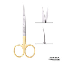
Considered the gold standard in bone grafting, an autograft is bone harvested from the patient's own body. Common intraoral donor sites include the chin and ramus. Because the material is the patient's own living tissue, it contains all three essential properties for bone regeneration: it is osteoconductive (acts as a scaffold), osteoinductive (signals new bone growth), and osteogenic (contains bone-forming cells). This unique combination results in the fastest and most predictable healing with virtually no risk of rejection or disease transmission. The harvesting procedure itself requires sharp, precise incisions, making reliable MacCut surgical blades an essential part of the instrument setup.
Allografts: Sourcing from a Human Donor
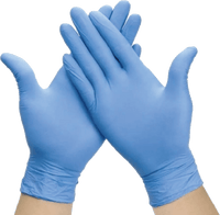
An allograft is a practical and widely used alternative sourced from a deceased human donor. This bone is processed in a tissue bank to ensure sterility and remove immunogenic components, making it a safe and effective option. Allografts are osteoconductive and, depending on the processing method (e.g., freeze-dried bone allograft or FDBA), can retain some osteoinductive properties. This eliminates the need for a second surgical donor site, reducing patient morbidity and surgical time. Since you are working with processed biological materials, maintaining a sterile field is paramount. Using high-quality MacSafe gloves and other infection control products is non-negotiable.
Xenografts: Sourcing from Animal Donors
Xenografts are derived from an animal source, most commonly bovine (cow) or porcine (pig). The bone is processed to remove all organic material, leaving behind a natural mineral scaffold that is primarily osteoconductive. Xenografts are excellent for maintaining space and providing long-term volume stability because they resorb very slowly. While they do not actively induce new bone formation, they provide a reliable framework for the patient's own bone cells to grow into. Placing and contouring this type of graft material often requires robust and ergonomic tools, such as the instruments found in our Hexa Series, which are designed for advanced surgical applications.
Alloplasts: Lab-Created Synthetic Grafts
Alloplasts are synthetic, biocompatible materials created in a laboratory. Common compositions include hydroxyapatite, tricalcium phosphate, and bioactive glass. These materials are purely osteoconductive, serving as a scaffold for new bone to infiltrate. The primary advantages of alloplasts are their unlimited supply, consistent quality, and zero risk of disease transmission. However, they lack any inherent biological activity to signal bone growth and are often considered when other options are not suitable. Regardless of the graft material, achieving primary closure over the site is crucial for success. Using the appropriate MacSuture product ensures the graft is protected and contained during the critical initial healing phase.
Why Autografts Are the Gold Standard
When it comes to bone grafting, autografts are widely considered the clinical gold standard. An autograft is bone harvested from one site in a patient’s body and transplanted to another. This method provides an ideal biological blueprint for regeneration because it contains the patient's own living cells, proteins, and minerals. The complete compatibility of the material means there is no risk of immune rejection, leading to highly predictable and successful integration.
While autografts offer unmatched regenerative properties, they require a second surgical site, which adds complexity to the treatment plan. As a clinician, it's essential to weigh the superior biological performance of autografts against the surgical demands and potential patient morbidity associated with the harvesting procedure. Understanding these factors will help you determine when this gold-standard approach is the right choice for your patient’s dental implant case.
How and Where to Harvest Autografts
The source of an autograft depends on the volume of bone required for the procedure. For smaller defects, intraoral sites are often preferred due to their proximity and ease of access. Common intraoral donor sites include the mandibular symphysis (chin), the external oblique ridge, and the ramus. For more extensive ridge augmentations requiring a larger volume of bone, extraoral sites like the iliac crest (hip) or tibia may be necessary. The harvesting technique requires precision, starting with a clean incision using a sterile surgical blade from the MacCut collection. Specialized instruments, like those in our ergonomic Hexa Series, are then used to carefully procure the bone block or particulate matter while minimizing trauma to the surrounding tissues.
Advantage: Unmatched Regenerative Potential
The primary advantage of an autograft lies in its biological properties. It is the only grafting material that possesses all three characteristics necessary for bone formation: it is osteogenic (contains living bone-forming cells), osteoinductive (contains growth factors that recruit and stimulate new bone growth), and osteoconductive (provides a natural scaffold for bone to grow upon). This powerful combination results in the most reliable and rapid healing. Because the graft is the patient’s own tissue, there is virtually no risk of rejection or disease transmission. This makes autografts the most effective option for promoting robust osseointegration and ensuring a stable, long-lasting foundation for dental implants.
Disadvantage: Requires a Second Surgical Site
The most significant drawback of using an autograft is the need for a second surgical site to harvest the bone. This additional procedure increases overall surgical time, complexity, and patient cost. The donor site also introduces a risk of morbidity, including postoperative pain, swelling, potential nerve damage, and scarring. The quantity of bone available from intraoral sites can be limited, which may not be sufficient for larger defects. Proper surgical technique and meticulous wound closure at both the donor and recipient sites, using reliable materials like MacSuture, are critical to minimizing these complications and ensuring a positive patient outcome.
Allografts: The Convenient Alternative to Autografts
When an autograft isn't a viable option, allografts present a highly effective and convenient alternative for bone augmentation. Sourced from human donors and processed by a tissue bank, these grafts eliminate the need for a second surgical site, which is a significant advantage for both you and your patient. This approach not only simplifies the procedure but also reduces patient morbidity and post-operative discomfort. Allografts are available in various forms, including mineralized and demineralized freeze-dried bone allograft (FDBA and DFDBA), giving you the flexibility to choose the best material for the specific clinical scenario.
These materials act as an osteoconductive scaffold, providing a natural framework that encourages the patient's own bone-forming cells to migrate and generate new bone. While they don't have the same osteoinductive potential as autografts, their proven reliability and off-the-shelf availability make them a cornerstone of modern implant dentistry. Proper handling and placement are key, and using precision instruments like those in our Hexa Series can ensure the graft is securely positioned for optimal integration. By understanding their processing and clinical behavior, you can confidently incorporate allografts into your practice to achieve predictable and successful outcomes.
How Allografts Are Processed for Safety
The safety of allograft material is paramount. To ensure patient safety, tissue banks follow stringent screening and sterilization protocols to eliminate the risk of disease transmission. The donated bone undergoes a rigorous processing regimen, which often includes freeze-drying and gamma irradiation, to render it sterile and reduce its antigenicity. This meticulous preparation minimizes the chance of an adverse immune response from the host. While these safety measures are essential, it's important to recognize that heavy processing can sometimes diminish the graft's inherent biological activity. Maintaining a sterile field with reliable infection control supplies remains a critical part of the procedure to support a complication-free healing environment.
Advantage: No Donor Site Surgery Required
The most compelling advantage of using an allograft is the complete avoidance of a donor site surgery. This immediately translates to a less invasive procedure, shorter operative time, and a more comfortable recovery for your patient. By eliminating the need to harvest bone from the iliac crest, tibia, or intraoral sites, you spare your patient from the pain, potential complications, and scarring associated with a second surgical area. This benefit often improves case acceptance, as patients are more receptive to a single-site surgery. Furthermore, allografts are readily available in unlimited quantities, providing a consistent and predictable supply for treating various defect sizes without limitations.
Disadvantages: Healing Variability and Cost
While allografts offer significant benefits, they also come with a few considerations. The healing process can be more variable and typically takes longer than with autografts, often requiring four to six months for complete integration. Because the material is from a different individual, there is a small, though rare, potential for an immune response or slower remodeling as the host body replaces the graft with its own bone. The extensive processing required to ensure safety can also reduce the graft's osteoinductive properties. Finally, the cost of processed allograft material can be higher than other options, which may be a factor in treatment planning and patient communication. Proper wound closure with quality materials like MacSuture is crucial to protect the graft and support predictable healing.
Common Forms: Putty, Paste, and Gels
Bone graft materials are available in several forms, each designed to simplify application and adapt to the specific surgical site. The most common options—putty, paste, and gel—offer distinct handling characteristics that can make a significant difference in your workflow. Bone graft putty is a malleable material that you can easily shape and pack into a defect, making it ideal for contouring and filling irregular voids in socket preservation or ridge augmentation. Pastes offer a more fluid consistency, allowing them to flow into smaller, hard-to-reach areas, which is especially useful in minimally invasive techniques. Finally, gels provide an injectable, viscous option that can be delivered with precision. The choice often comes down to the specific clinical need and your personal preference. Having versatile instruments, like those in the Pro Series, ensures you can confidently place any of these materials.
Xenografts: A Reliable Framework for New Bone Growth
When you need a grafting material that provides excellent space maintenance and a predictable scaffold for bone regeneration, xenografts are often the go-to solution. Derived from an animal source, these materials have become a cornerstone of regenerative dentistry, particularly for socket preservation, sinus lifts, and ridge augmentation. The key to their success lies in their processing. Through a highly controlled purification process, all organic and antigenic components are removed, leaving behind a natural, porous mineral matrix that is remarkably similar to human bone.
This structure acts as an ideal osteoconductive scaffold, meaning it provides the perfect framework for the patient’s own bone-forming cells to migrate into and begin depositing new bone. Because xenografts resorb very slowly, they offer exceptional volume stability over time, which is critical for preventing ridge collapse and ensuring an ideal foundation for future implant placement. For clinicians seeking a reliable and readily available option, high-quality bone grafting materials are essential for achieving consistent and successful clinical outcomes. The predictable nature of xenografts allows you to confidently plan your restorative cases from start to finish.
Comparing Bovine vs. Porcine Sources
Xenografts are sourced from non-human species, most commonly bovine (cow) or porcine (pig) bone. The raw material undergoes a rigorous deproteinization process that eliminates all organic matter, including cells, proteins, and potential pathogens. This critical step renders the material biocompatible and prevents an immune response from the patient. What remains is the inorganic, mineral component of the bone—primarily hydroxyapatite—with its natural micro- and macro-porosity intact. This preserved architecture is what allows it to function so effectively as a scaffold, guiding the infiltration of blood vessels and osteoblasts to build new, vital bone within the graft site.
Advantage: High Availability and Structural Integrity
One of the most significant advantages of xenografts is their unlimited availability, which eliminates the need for a second surgical site to harvest an autograft. This simplifies the surgical procedure for both you and the patient. Clinically, the primary benefit is the material’s structural integrity. Xenografts provide a stable, slow-resorbing scaffold that excels at maintaining the contour and volume of the alveolar ridge. This is especially crucial in aesthetic zones or in cases requiring significant augmentation before implant placement. The predictable space maintenance ensures that you have sufficient bone volume when you are ready to place the implant, contributing to long-term functional and aesthetic success. These materials are among the most well-documented bone substitutes in dentistry.
Disadvantages: Slower Remodeling and Immune Response Risks
While the slow resorption rate of xenografts is a major advantage for volume preservation, it can also be a clinical consideration. Because the material remodels slowly, particles of the graft may remain encapsulated within the new bone for years. The graft primarily serves as a scaffold rather than being fully replaced by the patient's own bone over time. Additionally, despite advanced purification methods that remove nearly all organic material, there remains a very low theoretical risk of an immune or inflammatory response. Proper patient selection and a thorough review of their medical history are important steps to mitigate any potential complications and ensure a predictable healing process.
Alloplasts: How Do Synthetic Grafts Compare?
Moving away from biologic sources, alloplasts represent the category of synthetic bone graft materials. These lab-engineered solutions are designed to provide a reliable, safe, and readily available alternative for ridge and socket preservation. Because they are man-made, they eliminate the risks associated with donor tissues and offer a high degree of predictability in their physical properties. For clinicians seeking a consistent and sterile scaffolding material, alloplasts present a compelling option in many dental implant cases.
What Are Synthetic Grafts Made Of?
Alloplasts are typically composed of biocompatible materials that mimic the mineral portion of natural bone. The most common formulations include hydroxyapatite (HA), tricalcium phosphate (TCP), and biphasic calcium phosphate (a combination of HA and TCP). These compounds are processed into granules, blocks, or putties that serve as an osteoconductive framework. This means they create a stable, porous scaffold that encourages the patient’s own bone-forming cells to migrate into the defect and generate new bone over time, effectively using the body's natural healing potential to rebuild the site.
Advantage: Exceptional Safety and Predictable Results
One of the greatest advantages of alloplasts is their impeccable safety profile. Since they are entirely synthetic, there is zero risk of disease transmission from a donor. This eliminates patient concerns and complex consent discussions. Furthermore, their unlimited supply and off-the-shelf availability make them a convenient choice for any practice. The manufacturing process allows for precise control over particle size, porosity, and resorption rate, ensuring a consistent product from one case to the next. This predictability is invaluable when performing advanced procedures like implant preparations, where precise Hexa Series instruments and reliable materials are essential for success.
Disadvantage: Lacks Natural Biological Activity
The primary limitation of alloplasts is their lack of osteoinductive properties. Unlike autografts, they do not contain growth factors or viable cells to actively signal new bone formation. Their function is purely passive scaffolding. This can lead to a slower healing and integration process compared to biologic grafts. Resorption rates can also be a factor; some synthetic materials are designed to resorb slowly or not at all, which may not be ideal for every clinical scenario. Meticulous surgical technique and proper wound closure using high-quality MacSuture are critical to protect the graft and support the body’s slower regenerative response.
Limitations in Large Defects
While all graft materials perform well in contained, smaller defects, their limitations become more apparent as the size of the required augmentation increases. Large defects present a significant biological challenge because they require more than just a scaffold; they need powerful signaling to recruit blood vessels and bone-forming cells across a large, avascular space. Off-the-shelf materials like allografts, xenografts, and alloplasts are primarily osteoconductive. In a large defect, their lack of robust osteoinductive properties can lead to slower, less predictable healing or incomplete bone fill. The slow remodeling of xenografts and the biological inertness of alloplasts are particularly challenging in these scenarios, where active regeneration is critical. Managing these complex cases requires exceptional surgical skill and instrumentation, which is why many clinicians turn to tools like the Hexa Series, designed for precision in demanding procedures.
Even the gold-standard autograft has practical limitations when it comes to large defects. While it provides the ideal combination of osteogenic cells and osteoinductive growth factors, harvesting a sufficient volume of bone often requires an extraoral donor site, such as the iliac crest. This significantly increases the surgical complexity, operative time, and potential for patient morbidity, including pain and scarring at the harvest site. This trade-off often makes it a less appealing option for both the patient and the clinician, despite its superior biological performance. Regardless of the material chosen, protecting the graft is paramount, and achieving tension-free primary closure with a durable material like MacSuture is essential for a successful outcome.
How to Choose the Right Graft Material for Your Patient
Selecting the ideal bone graft material isn’t about finding a single "best" option—it's about identifying the right material for a specific patient and a unique clinical scenario. The decision is a careful balance of science, surgical goals, and patient-specific factors. Your clinical judgment is paramount, but it’s guided by three core considerations: the nature of the defect, the patient's overall health, and the logistics of the treatment plan.
Think of it as creating a customized treatment strategy. A young, healthy patient with a small, contained alveolar ridge defect will have different needs than an older patient with systemic health issues requiring a significant sinus lift. The amount of bone needed, the patient's ability to heal, and your own surgical preferences all come into play. By systematically working through these key areas, you can confidently choose a material that provides a stable foundation for implant success and aligns with your patient's long-term oral health goals. This thoughtful approach ensures predictable outcomes and reinforces the trust your patients place in your care.
Assess the Defect's Size and Location
The anatomy of the bone defect is the first piece of the puzzle. The sheer volume of bone needed will immediately narrow your options. For extensive ridge augmentations or significant vertical defects, an autograft harvested from a secondary site might be the only choice that provides sufficient volume and the necessary osteoinductive properties. For smaller, more contained defects, such as socket preservation, a xenograft or alloplast can provide an excellent osteoconductive scaffold without the morbidity of a second surgical site.
The location also matters. A sinus augmentation procedure often benefits from a particulate graft that is easy to pack and contains, while a block graft might be better suited for horizontal ridge augmentation. Your choice of surgical instruments and technique will also be tailored to the defect, ensuring you can properly prepare the site and stabilize the chosen material for optimal healing.
Evaluate Your Patient's Overall Health
A graft material is only as good as the patient's ability to heal around it. A thorough review of your patient's medical history is non-negotiable, as systemic factors can significantly impact osseointegration. As one study notes, the best choice depends on factors like "your health... and your personal concerns." Be particularly mindful of conditions that compromise vascularity and immune response.
Patients with uncontrolled diabetes, autoimmune diseases, or a history of heavy smoking may have impaired healing, making them higher-risk candidates. Similarly, those taking certain medications, like bisphosphonates, require careful evaluation. For these patients, a highly biocompatible material with strong regenerative potential may be necessary. Ensuring a sterile field with proper infection control protocols is always critical, but it becomes even more important when a patient's healing capacity is a concern.
Consider Surgical Complexity and Timelines
Your treatment plan's timeline and complexity are final critical factors. Are you planning a staged approach or placing the implant at the same time as the graft? The answer influences which material is most appropriate. Bone grafts typically require four to six months of healing before an implant can be placed, but this can vary based on the material and the size of the graft.
For complex cases requiring significant structural support over a longer healing period, a slow-resorbing xenograft might be ideal, as it maintains space and acts as a durable scaffold. For simpler cases where faster bone turnover is desired, an allograft or certain alloplasts might be more suitable. Discussing these timelines with your patient manages their expectations and ensures they are committed to the full treatment process, from the initial graft to the final restoration.
Healing Timelines and Outcomes: What to Expect
Understanding the healing trajectory of different bone graft materials is fundamental to successful treatment planning and managing patient expectations. The time from graft placement to implant readiness isn't just a waiting period; it's a critical phase of biological integration that determines the long-term stability of the final restoration. While every patient's healing capacity is unique, the choice of graft material sets a predictable timeline for osseointegration. This allows you to map out a clear surgical plan, from the initial augmentation procedure to the final implant placement. By mastering these timelines and the factors that influence them, you can confidently guide your patients through the process and ensure the augmented site is perfectly prepared for a stable, lasting implant.
The Surgical Procedure: A Step-by-Step Overview
Once you've selected the appropriate graft material, the success of the procedure shifts to your surgical technique. Meticulous execution at every stage—from site preparation to final closure—is what protects the graft, promotes vascularization, and ultimately determines the quality of the regenerated bone. A successful outcome depends on creating an ideal biological environment where the body can do its work, and that starts with a clean, precise, and well-managed surgical field.
Preparing the Site and Placing the Graft
The first surgical step is to prepare a clean, bleeding, and well-vascularized recipient bed. This is crucial because the graft material relies on a healthy blood supply to initiate the healing cascade. After reflecting the flap, you must thoroughly debride the defect of any soft tissue remnants to ensure direct contact between the graft and the host bone. The graft material is then carefully placed and contoured to fill the defect and restore the desired ridge anatomy. This step requires both precision and control, which is why many clinicians prefer the ergonomic, non-slip grip of instruments from the Hexa Series. These tools are specifically designed for advanced surgical procedures, ensuring you can manipulate and compact the graft material effectively for a stable result.
Wound Closure and Protection
Proper wound closure is arguably one of the most critical steps for a predictable outcome. The goal is to achieve a tension-free primary closure that completely isolates the graft from the oral environment. This protective seal prevents bacterial contamination, contains the graft material, and stabilizes the blood clot, which is essential for initiating angiogenesis and bone formation. Using a high-quality, biocompatible suture is non-negotiable. The right suture material, like our reliable MacSuture products, allows you to secure the flap without compromising blood flow to the tissue margins. This meticulous closure protects your hard work and provides the undisturbed healing environment necessary for successful bone regeneration.
How Graft Integration Varies by Material
The type of graft material you select directly influences the healing timeline and when you can schedule implant placement. Generally, you can expect a healing window of four to six months, but this varies significantly. Autografts, containing the patient's own living cells, integrate the fastest, typically ready for an implant in about three to four months. Allografts follow, requiring around four to six months for proper integration. Xenografts, with their slower resorption rate, often need a longer period of six to nine months. Finally, alloplastic materials can have the most variable and extended timelines, sometimes taking six to twelve months to achieve the necessary stability for implant loading.
What Are the Keys to Successful Osseointegration?
A successful bone graft does more than just fill a void; it actively rebuilds the foundation for an implant. The ideal material should excel in four key biological processes. It must achieve osseointegration, bonding securely to the host bone. It should also promote osteogenesis, or the formation of new bone from cells within the graft material itself. Furthermore, it needs to be osteoconductive, providing a stable scaffold for the patient's own bone cells to grow into. Finally, osteoinduction is crucial, as it signals the body’s stem cells to differentiate into bone-forming cells. Achieving these outcomes relies heavily on precise surgical technique, where clean incisions made with a sharp MacCut surgical blade can minimize tissue trauma and support optimal healing.
How to Prepare the Graft Site for Implant Placement
Thanks to modern advancements in bone grafting, more patients than ever are candidates for dental implants. The ultimate goal of grafting is to create a dense, vascularized, and dimensionally stable site that can support an implant for years to come. Before proceeding with placement, it's essential to verify that the graft has fully integrated and matured into solid bone. Proper site preparation and meticulous surgical closure are paramount for protecting the graft during the initial healing phase. Using high-quality sutures, such as those from the MacSuture collection, ensures the soft tissue is securely closed, preventing contamination and promoting an undisturbed environment for bone regeneration. This careful preparation paves the way for a predictable and successful implant procedure.
Post-Operative Care and Patient Guidance
The success of a bone graft doesn't end when the patient leaves the chair. Clear, concise post-operative instructions are just as critical as the surgical procedure itself. When patients understand what to do and what to expect, they become active partners in their own healing. Providing them with detailed guidance on managing discomfort, diet, and activity not only improves clinical outcomes but also reduces post-op complications and anxious phone calls. This is your opportunity to reinforce their trust and ensure the graft site remains undisturbed during the most critical phase of healing.
Managing Swelling and Discomfort
Setting realistic expectations about swelling and discomfort is key to a positive patient experience. Inform patients that swelling is a normal part of the healing process and will likely peak two to three days after the procedure. As one clinic advises, recommend applying an ice pack to the cheek over the surgical area for the first 48 hours, using a cycle of 20 minutes on and 20 minutes off to help minimize inflammation. For pain management, provide clear instructions for any prescribed medications or recommend an appropriate over-the-counter regimen. A well-prepared post-op kit, including sterile gauze and written instructions, can make a significant difference in patient compliance and comfort.
Dietary and Activity Restrictions
Protecting the graft site from physical disruption is paramount, and dietary instructions are your first line of defense. Advise patients to stick to cool, soft foods for the first 24 hours and to avoid anything hot, spicy, or crunchy that could irritate the wound. Crucially, instruct them not to spit or use a straw for at least two days, as the negative pressure can dislodge the blood clot essential for healing. It's also wise to recommend they avoid strenuous physical activity for several days, as an elevated heart rate can increase bleeding and swelling at the surgical site. These simple precautions help ensure the graft remains stable and protected.
Managing Patient Expectations During Recovery
The healing journey for a bone graft is a marathon, not a sprint. Effectively managing patient expectations from the outset prevents confusion and anxiety down the road. It’s important to clearly communicate the difference between normal post-operative symptoms and the warning signs of a potential complication. By providing a clear roadmap of the full healing timeline—from the initial soft tissue recovery to the months-long process of bone integration—you empower patients with the knowledge they need to navigate their recovery with confidence. This proactive communication strengthens the doctor-patient relationship and ensures a smoother path to the final implant restoration.
Normal Healing vs. Signs of Complications
Educating patients on what to expect can prevent unnecessary worry and ensure they know when to seek help. Normal healing includes manageable pain, some swelling and bruising, and minor bleeding in the first 24 hours. In contrast, signs of a potential complication or graft failure include pain or swelling that worsens after the first week, the presence of pus, or the feeling that the gums are pulling away from the teeth. Providing a printed handout that clearly lists these differences gives patients a reliable reference. This simple tool helps them distinguish between a normal recovery and a situation that requires your immediate attention, ensuring any issues are addressed promptly.
The Full Healing Timeline: From Weeks to Months
It's crucial to explain that there are two distinct phases of healing. The initial soft tissue healing, where tenderness and swelling subside, typically takes about one to two weeks. Patients will feel significantly better during this time, but it's vital they understand that the real work is happening beneath the surface. The second phase, bone integration, is a much longer process. As the Cleveland Clinic notes, the graft needs anywhere from three to twelve months to fully mature and become strong enough to support an implant. This extended timeline is necessary for the graft to be replaced by the patient's own strong, vital bone, creating the solid foundation required for long-term implant success.
Managing Potential Risks and Complications
While bone grafting is a highly predictable procedure with excellent success rates, no surgical intervention is entirely without risk. Proactive management begins with understanding the potential complications, recognizing early warning signs, and educating your patients on what to expect during the healing process. A well-informed patient is your best partner in identifying issues before they escalate. By preparing for these possibilities and ensuring your practice is equipped with the necessary tools and materials for any scenario, you can confidently handle challenges as they arise, safeguarding both the patient's health and the long-term success of the implant.
Common Complications to Monitor
Following a bone grafting procedure, it's important to monitor for several common complications. Post-operative infection is a primary concern, though its risk can be significantly minimized with a strict aseptic technique and the use of reliable infection control supplies. Swelling, mild pain, and bruising are normal physiological responses but should progressively decrease after the first few days. You should also watch for graft migration or instability, where the particulate material shifts from its intended position. In maxillary procedures, sinus membrane perforation is a known risk, with some studies reporting its occurrence in a significant percentage of cases. While most of these issues are manageable, they require careful observation during follow-up appointments to ensure a smooth and uneventful recovery for your patient.
Recognizing Signs of Graft Failure
Distinguishing between normal post-operative healing and the early signs of graft failure is a critical clinical skill. While some initial discomfort is expected, pain or swelling that worsens after the first week is a significant red flag and may indicate an underlying infection or inflammatory response. The presence of purulent exudate from the surgical site is a definitive sign of infection that requires immediate intervention. Other indicators of failure include gingival recession that exposes the graft material, a palpable lack of firmness at the site after several weeks, or radiographic evidence showing no new bone formation. Educating patients to report these symptoms promptly allows for timely management and can sometimes salvage a failing graft.
Specific Risks in Sinus Lift Procedures
Sinus augmentation procedures carry their own unique set of risks, with perforation of the Schneiderian membrane being the most common intraoperative complication. This delicate membrane can be easily torn during sinus floor elevation, potentially leading to graft displacement into the sinus cavity and subsequent infection or failure. Managing a perforation requires skill and the right equipment; small tears can often be repaired with a collagen membrane, but larger ones may necessitate aborting the procedure. The precision and control offered by specialized instruments, like those in the Hexa Series, are invaluable for these delicate maneuvers. Post-operatively, patients should also be monitored for signs of acute sinusitis, which can arise if the sinus ostium becomes blocked.
Key Considerations for Your Dental Practice
Selecting the right bone graft material is a decision that balances clinical evidence, patient factors, and practical considerations. Once you’ve assessed the defect and your patient’s health, a few final points can help guide your choice to ensure a predictable and successful outcome. These factors often involve the tactile experience in the operatory, transparent conversations with your patient, and a clear understanding of long-term performance.
Evaluate Handling Characteristics and Biocompatibility
The ideal bone graft material should not only be biocompatible but also easy to handle. In the midst of a procedure, you need a material that packs well, stays where you place it, and doesn’t wash out. Autografts are the benchmark for biocompatibility since the tissue is the patient’s own, eliminating any risk of rejection. However, the need for a second surgical site is a significant drawback. Allografts and synthetic materials offer excellent off-the-shelf convenience and eliminate donor site morbidity. Your ability to precisely place and contour the graft is supported by your instrumentation, where tools from the Hexa Series provide the ergonomic control needed for such delicate work.
How to Balance Cost with Patient Expectations
Cost is an unavoidable part of any treatment plan, and it’s important to discuss it openly with your patients. Autografts are typically the most expensive option because they require a secondary surgical procedure and potentially a longer time in the operating room. Allografts, xenografts, and alloplasts are often more affordable and can provide excellent outcomes without the discomfort and recovery associated with a donor site. Clearly explaining the trade-offs between each material—balancing cost, procedural complexity, and healing potential—empowers patients to make an informed choice. When patients understand the value and rationale behind your recommendation, they are more likely to move forward with confidence.
Comparing Material Costs
While clinical suitability always comes first, the financial aspect of bone grafting is a practical reality for every patient. Generally, synthetic alloplasts and animal-derived xenografts are the most affordable materials, with a typical cost between $300 and $800. Allografts, sourced from human donors and subject to extensive processing, carry a higher price tag, with patients often seeing a cost between $700 and $1,500. Although the autograft material itself has no direct cost, the second surgical procedure—factoring in anesthesia and operating room time—makes it the most expensive option overall. Walking your patients through these differences helps them understand the value behind your recommendation, which is key for building trust and improving case acceptance.
Understanding Long-Term Success Rates
Ultimately, the goal is successful osseointegration that provides a stable foundation for a future implant. Clinical studies show that autografts, allografts, and xenografts can all achieve very high long-term success rates. The key difference often lies in the healing timeline, which typically ranges from four to six months before the site is ready for implant placement. Setting realistic expectations about this timeline is crucial for patient satisfaction. Success isn't just about the material; it depends on meticulous technique and proper post-operative care. Ensuring secure wound closure with high-quality MacSuture products protects the graft during the critical early stages of healing, paving the way for predictable integration.
Frequently Asked Questions
Related Articles
Tags
Blog posts
-

, by SurgiMac How to Disinfect with Metrex CaviWipes Correctly
-
, by SurgiMac Suture Sizes From Smallest to Largest: A Complete Guide
-

, by Marketing SurgiMac Stop the Bleeding: 5 Procurement Leaks Draining DSO Margins in 2026


