
Dental Surgical Instruments for Extractions
- , by SurgiMac
- 13 min reading time
Compare dental surgical instruments for access, luxation, delivery, and closure, with practical selection guidance for extraction trays.
Dental surgical instruments directly influence access, control, and tissue management during an extraction. For professional dental teams, the right tray supports deliberate socket expansion, secure tooth delivery, and efficient closure without substituting force for technique. SurgiMac helps U.S. practices source dependable instruments for each procedural phase, from the initial incision through postoperative site management. This guide explains how to select, organize, inspect, and maintain an extraction set according to anatomy, clinical objectives, and reprocessing requirements.
Explore SurgiMac dental instruments for a procedure-ready extraction tray.
SurgiMac recommends building an extraction tray by procedural role: access and visualization, luxation, delivery, debridement, and closure. The appropriate dental surgical instruments depend on tooth position, root morphology, periodontal support, restorative condition, surgical access, and the clinician's planned technique. A role-based setup gives the assistant a predictable sequence while ensuring that the operator can escalate from routine elevation to surgical access without avoidable interruptions.
What dental surgical instruments belong on an extraction tray?
A complete tray is not simply a large assortment of instruments. It is a deliberately selected system that supports the planned extraction and foreseeable contingencies. Review the clinical examination and diagnostic imaging before setup.
Root curvature, divergence, hypercementosis, previous endodontic treatment, extensive caries, ankylosis risk, and proximity to adjacent anatomy may all change which instruments should be immediately available.
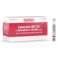
For routine cases, begin with an examination set, local anesthetic setup, suction, a periosteal elevator, suitable elevators or luxators, and forceps matched to the tooth and arch. Add a curette and irrigation setup for socket management. If flap elevation, sectioning, bone removal, or primary closure may be required, prepare the appropriate surgical handpiece and burs, scalpel, retractors, tissue forceps, needle holder, scissors, and suture material before the procedure begins.
| Procedural role | Common instrument choices | Primary selection question | Inspection priority |
|---|---|---|---|
| Access and visualization | Scalpel, periosteal elevator, retractor, suction | Will a flap be required, and which anatomy must remain visible? | Blade security, tip condition, retractor edges |
| Luxation | Straight elevator, apical elevator, luxator | Which tip geometry can enter the available periodontal ligament space? | Straight shaft, intact and sharp working end |
| Delivery | Universal or tooth-specific forceps | Do the beaks adapt apically to the root morphology? | Beak alignment, hinge movement, handle grip |
| Socket management | Curette, irrigation syringe, suction | What debridement is clinically indicated? | Curette edge, syringe and suction integrity |
| Closure | Needle holder, tissue forceps, scissors, sutures | Which needle and suture support the flap design and tissue tension? | Jaw alignment, ratchet, scissor edge |

How should dentists select forceps for extraction?
Forceps selection begins with anatomy rather than habit. The beaks should adapt to the cervical root surface as apically as practical, while the handle and beak angulation should provide access without requiring an unstable wrist position. Maxillary forceps commonly align differently from mandibular forceps because the operator approaches each arch from a different direction. Molar forceps may also have right- and left-specific designs that engage furcation anatomy.
Choose between universal and tooth-specific forceps according to the procedure and the range of cases treated. Universal patterns can make a routine setup efficient, but a tooth-specific pattern may improve adaptation when root morphology is complex. Cowhorn designs can engage the furcation of selected mandibular molars. Bayonet-style profiles can improve access in specific maxillary situations. The presence of a crown does not eliminate the need to understand the root form below it.
During evaluation, confirm that the beaks meet as designed, the hinge moves smoothly, and the handle texture supports controlled grip with gloved hands. Excessive play at the joint or misaligned beaks can compromise tactile feedback and force distribution.
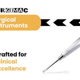
SurgiMac instrument lines, including lightweight and slim-profile options, allow teams to compare balance, access, and handle geometry while maintaining a professional extraction setup.
When should you use elevators or luxators?
Elevators and luxators serve related but distinct functions. Elevators commonly apply controlled rotational, wedging, or lever action to initiate mobility and expand the socket. Luxators generally have thinner, sharper blades designed to enter the periodontal ligament space and sever fibers with carefully directed apical pressure. Both require a stable grasp, clear visualization, and a planned direction of force.
Select tip width and curvature according to the available access and root surface. A straight elevator can be useful for initial mobility, while an apical elevator may provide access to a retained root fragment when used with an appropriate purchase point. Curved profiles can improve access in posterior regions, but their shape also changes the direction in which force is transmitted. The clinician should anticipate the path of movement before engaging the instrument.
Controlled technique protects the alveolar housing and adjacent structures. Avoid using an adjacent tooth as a fulcrum, and do not advance a working end without knowing where it will travel if resistance suddenly decreases. If elevation is not producing the expected mobility, reassess the root anatomy, access, and need for sectioning or bone removal rather than increasing force without a clear endpoint.
Compare SurgiMac instruments by access, handle design, and procedural role.
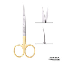
What blades and access instruments support precise surgery?
When surgical access is indicated, a sharp blade and stable handle support a deliberate incision with clean margins. A number 15 blade is commonly used for intraoral access because its compact curved edge permits controlled incisions around teeth and edentulous ridges. A number 12 blade may be useful for selected posterior or mucogingival applications. The blade profile should match the incision design, access angle, and operator preference.
Blade sharpness matters because a compromised edge can require additional pressure and reduce incision control. Use a new sterile blade according to office protocol, secure it correctly on a compatible handle, and manage placement and removal with an established sharps procedure. SurgiMac's MacCut surgical blades give practices an option for stocking procedure-specific blade profiles alongside their reusable instrumentation.
After incision, a periosteal elevator should reflect the flap while maintaining close contact with bone. Retractors then maintain access and protect soft tissue, but they should not create unnecessary pressure at the flap margin. Suction selection also matters: the tip must preserve visualization without repeatedly engaging delicate tissue. Together, these instruments create an operative field in which the clinician can see anatomy and direct each subsequent movement.
For SurgiMac customers, an effective access set combines a secure scalpel handle and appropriate blade with a periosteal elevator, retractor, and surgical suction. Each component should support visibility and controlled tissue handling. Access instruments should be selected as a coordinated group, not as isolated products, because poor visibility can compromise otherwise appropriate elevation and delivery technique.
How should a team coordinate closure instruments?
Closure begins before the first incision, with a flap design and tissue-management plan that anticipate repositioning. A basic closure set usually includes a needle holder, tissue forceps, scissors, and the selected suture. The needle holder jaws must securely engage the needle without twisting it, and the instrument size should match the needle and working area. Castroviejo-style holders can support fine control in selected procedures, while ring-handled holders remain familiar for many routine closures.
Tissue forceps should stabilize the flap without crushing it. Toothed tips may engage keratinized tissue with limited pressure, while other tip designs may be appropriate for more delicate tissue. Scissors must cut cleanly near the knot without fraying the suture. Review SurgiMac's guide to needle holder types and uses when standardizing closure sets across operators.
Suture selection should reflect tissue tension, expected healing, contamination risk, clinician preference, and whether removal is desirable. Absorbable and nonabsorbable materials each have appropriate uses, and needle curvature influences access. Teams should follow the manufacturer's instructions for handling and storage. The goal is stable approximation without excessive tension, not simply rapid completion of the final procedural phase.
How do you evaluate quality, ergonomics, and reprocessing fit?
Instrument value includes performance throughout its service life, not only purchase price. Evaluate steel quality, finish, joint construction, tip integrity, resistance to corrosion, and the manufacturer's care instructions. A durable instrument should retain its intended geometry through repeated clinical use and validated reprocessing. Instruments with damaged working ends or unreliable joints should be removed from service rather than retained as backups.
Ergonomics affect clinical control and occupational comfort. Handle diameter, texture, weight, and balance influence how securely an operator can guide the working end. Slim profiles may improve access, while lightweight handles may reduce cumulative hand fatigue. The best choice remains operator- and procedure-dependent, so practices should assess instruments with the gloves and techniques used in their normal workflow.
Confirm that every reusable instrument is compatible with the practice's cleaning, packaging, sterilization, and storage procedures. Follow the instrument manufacturer's instructions for use and the practice's infection-prevention protocol. Hinged instruments may need to be processed in an open position, and joints require inspection for retained debris and smooth movement. Do not allow saline or organic material to dry on instruments before cleaning.
- Inspect tips, cutting edges, hinges, ratchets, beaks, and handles during tray assembly.
- Remove instruments with corrosion, cracks, pitting, looseness, bending, or misalignment.
- Use cassettes or protective systems that reduce working-end damage during processing.
- Keep procedure sets standardized, while allowing documented variations for clinician preference.
- Track repairs and replacements so recurring failures can inform purchasing decisions.
What should clinicians verify before each extraction?
A preprocedural verification should connect the treatment plan to the instruments on the tray. Confirm the patient, tooth, planned procedure, current imaging, medical considerations, and consent according to practice policy. The clinician and assistant should then review whether routine delivery is expected or whether surgical access, sectioning, root retrieval, or closure may be required. This brief discussion allows the team to prepare appropriate dental surgical instruments before treatment rather than responding to a predictable need after the procedure is underway.
Verify that tooth-specific forceps and intended elevators are present and serviceable. If a flap is possible, confirm that the scalpel handle, selected blade, periosteal elevator, retractor, suction, and closure module are available. When sectioning or bone removal is planned, check the surgical handpiece, bur selection, irrigation, and suction before local anesthetic administration. The assistant should also confirm that packaged items are intact and within the manufacturer's labeled use period.
Instrument readiness does not determine the treatment plan. If clinical findings differ from the examination or imaging, pause and reassess rather than continuing with an unsuitable setup or technique. Additional imaging, referral, or a revised surgical approach may be indicated. A well-organized tray supports that decision-making by making instrument limitations visible early and reducing pressure to proceed simply because a case has started.
After extraction, account for sharps and instruments, inspect the site according to the clinical plan, and transfer reusable items into the established reprocessing workflow. Document any broken instrument, damaged tip, or unexpected difficulty that could affect follow-up care or future tray design. These steps connect chairside performance with quality assurance and help the practice identify recurring equipment or workflow issues.
How can practices standardize extraction trays?
Standardization improves readiness without making every case identical. Create a core extraction cassette for frequently used instruments, then define add-on modules for surgical access, retained roots, molar delivery, and closure. This approach helps assistants identify missing items quickly and limits unnecessary instruments that would otherwise require reprocessing after the procedure.
Document the expected tray layout with a photograph or diagram, and organize instruments in the sequence in which they are likely to be used. Before seating the patient, the team should confirm imaging, planned procedure, required instruments, and likely escalation needs. After the case, record instrument failures or workflow gaps while details are fresh. Periodic review turns those observations into a more reliable setup.
SurgiMac supports extraction-tray standardization by helping professional dental teams source instruments by function and compare designs for access, delivery, and closure. A standardized core set, combined with case-specific modules, improves procedural readiness while keeping inventory and reprocessing demands manageable.
Practices can also consolidate purchasing through a dependable supplier while retaining clinical choice. Explore SurgiMac for professional dental and surgical supplies, then review product specifications and care instructions before adding an instrument to a standardized set. Purchasing decisions should account for expected case volume, operator preferences, replacement availability, and compatibility with existing cassettes.
- Review examination findings and diagnostic imaging before selecting the core set.
- Arrange instruments by access, luxation, delivery, socket management, and closure.
- Add case-specific modules for retained roots, surgical access, or primary closure.
- Verify instrument condition and reprocessing compatibility during tray assembly.
- Document workflow gaps after the procedure and refine the standardized set.
Build or refine your extraction setup with SurgiMac dental instruments.
Frequently asked questions
What instruments are essential for a routine dental extraction?
A routine extraction tray generally includes an examination set, periosteal elevator, appropriate straight and apical elevators, tooth-specific or universal forceps, curette, suction, and instruments for closure when indicated. The final selection should reflect the tooth, root morphology, access, imaging findings, and the clinician's planned technique.
How do elevators differ from luxators?
Elevators are commonly used to apply controlled leverage and expand the socket, while luxators have thinner, sharper blades intended to enter the periodontal ligament space and sever fibers. Neither should be used with uncontrolled force, and adjacent teeth should not serve as fulcrums.
How often should extraction instruments be inspected?
Dental teams should inspect instruments during tray assembly and again after reprocessing. Remove an instrument from service if it has corrosion, a loose joint, a bent or damaged tip, misaligned beaks, a compromised cutting edge, or a ratchet that does not engage consistently.
Which scalpel blade is commonly used for oral surgical access?
A number 15 blade is commonly selected for many intraoral incisions because its small curved cutting edge supports controlled access. A number 12 or other blade profile may be preferred for specific posterior or mucogingival access, according to the clinician's technique.
Related Articles
Tags
Blog posts
-

, by SurgiMac How to Disinfect with Metrex CaviWipes Correctly
-
, by SurgiMac Suture Sizes From Smallest to Largest: A Complete Guide
-

, by Marketing SurgiMac Stop the Bleeding: 5 Procurement Leaks Draining DSO Margins in 2026


